|
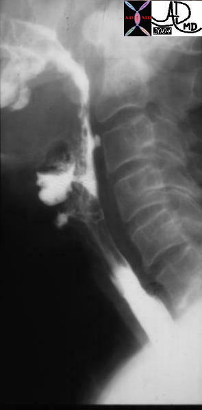
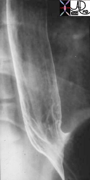
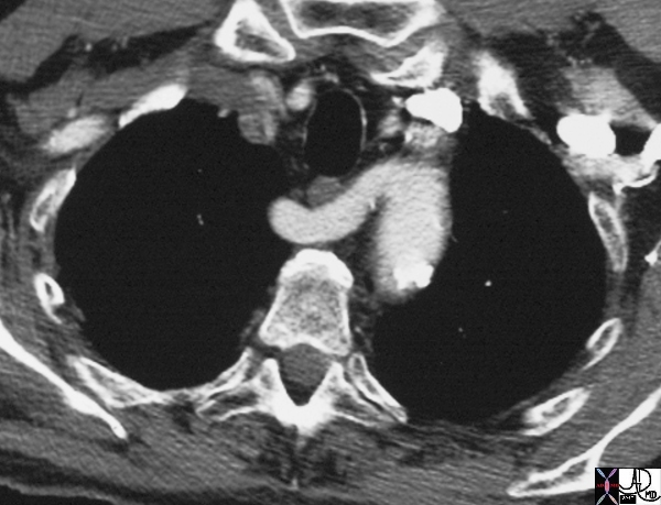
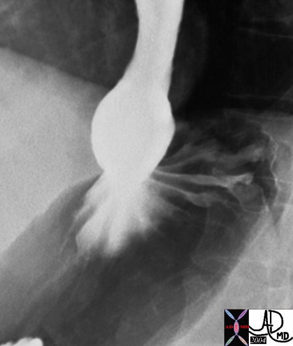
Position Copyright 2007 The esophagus extends downward through the lower portion of the neck and the superior and posterior mediastina of the thorax. It then passes through the esophageal hiatus of the diaphragm to join the cardia of the stomach at about the level of the tenth thoracic vertebra. It is found in both the thoracic cavity and to a much lesser extent in the abdominal cavity. In the chest it is part of the mediastinum and forms a gentle reversed “S” shape around the midline. Craniocaudal positioning: cranially it is slightly to the right and caudally slightly to the left. Anteroposterior positioning: as a relatively posterior structure it lies in close contact with the vertebral bodies. The cervical portion originates at about C6 opposite the cricoid cartilage, and lies within the lower portion of the neck and projects approximately 1/4″ to the left of the tracheal margin. The thoracic portion lies in the superior and posterior mediastinum. It is a midline structure at the level of T4 behind the aortic arch. It has an inclination to the right until about the level of T7. At about the level of the 10th thoracic vertebra, it again turns left more sharply than in its previous curves and passes through the esophageal hiatus. It travels behind the aortic arch and the left main stem bronchus. Low in the thorax, the esophagus passes anterior to the aorta and enters the abdomen, slightly to the left of midline, through the esophageal hiatus of the diaphragm. The abdominal portion resides in the upper abdomen for a short distance before it connects with the stomach at the GE junction.
Relations: Anterior to the cervical portion lies the membranous wall of the trachea, to which it is rather loosely connected by areolar tissue and some muscular strands, so that the anterior esophageal and the posterior tracheal walls are occasionally referred to as the “common party wall.”
In the groove on each side between the trachea and the esophagus ascend the recurrent laryngeal nerves. Posteriorly, the esophagus lies here upon the vertebral bodies and thus longus colli muscles, with the prevertebral fascia intervening. On each side the carotid sheath and the structures it contains accompany the cervical esophagus. Owing to the aforementioned curvature of the esophageal tube to the left in this region, it is closer to the carotid sheath on this side than it is on the right. The lobes of the thyroid gland partially overly the esophagus on each side. The thoracic duct ascends in the root of the neck on the left side of the esophagus and then arches laterally behind the carotid sheath and anterior to the vertebral vessels to enter the left brachiocephalic or left subclavian vein at the medial margin of the anterior scalene muscle.
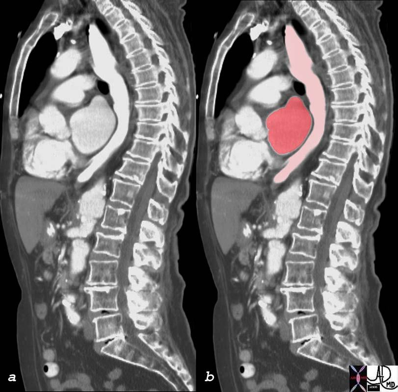
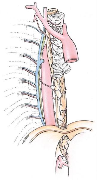
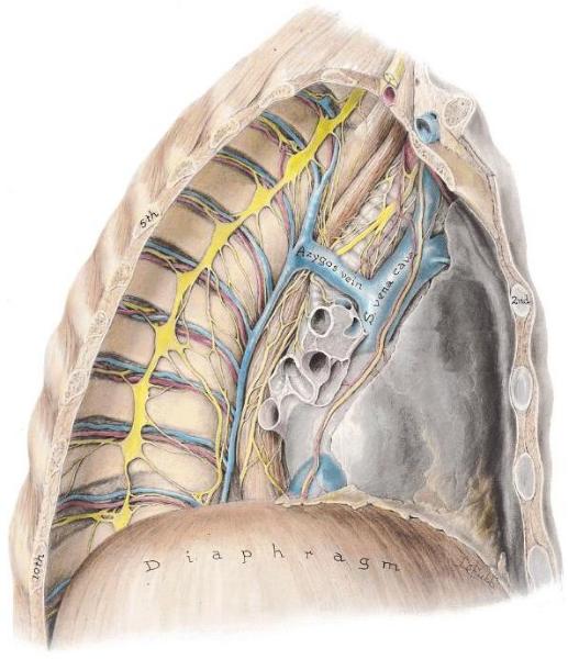
The thoracic esophagus continues to lie posterior to the trachea as far as the level of the fifth thoracic vertebral body, where the trachea bifurcates. The trachea deviates slightly to the right at its lower end, so that the left main bronchus crosses in front of the esophagus. Below this point the esophagus is separated anteriorly from the left atrium of the heart by the pericardium. In the very lowest portion of its thoracic course, the esophagus passes behind the diaphragm to reach the esophageal hiatus. On the left side the esophageal wall in the upper thoracic region is the ascending portion of the left subclavian artery and the parietal pleura at about the level of the fourth thoracic vertebra, the arch of the aorta passes backward and alongside the esophagus. Below this point the descending aorta lies to the left, but when that a vessel passes behind the esophagus, the left mediastinal pleura again comes to adjoin the esophageal wall. On the right side the right parietal pleura is intimately applied to the esophagus, except when, at about the level of the fourth thoracic vertebra, the azygos vein intervenes as it turns forward. In the upper part of its thoracic course, the esophagus continues to lie upon the longus colli muscle and the vertebral bodies, with the prevertebral fascia intervening. At about the level of the eighth thoracic vertebra, however, the aorta comes to lie behind the esophagus. The azygos vein ascends behind and to the right of the esophagus as far as the level of the fourth vertebral body, where it turns forward. The hemiazygos vein also crosses from left to right behind the esophagus, as do the upper five right intercostal arteries. The thoracic duct, ascending first to the right of the esophagus, inclines to the left behind it at about the level of the fifth vertebral body, to continue its ascent on the left side of the esophagus. In its short abdominal portion the esophagus lies upon the diaphragm, with the esophageal impression of the liver applied tunnel like to its anterior aspect.
|