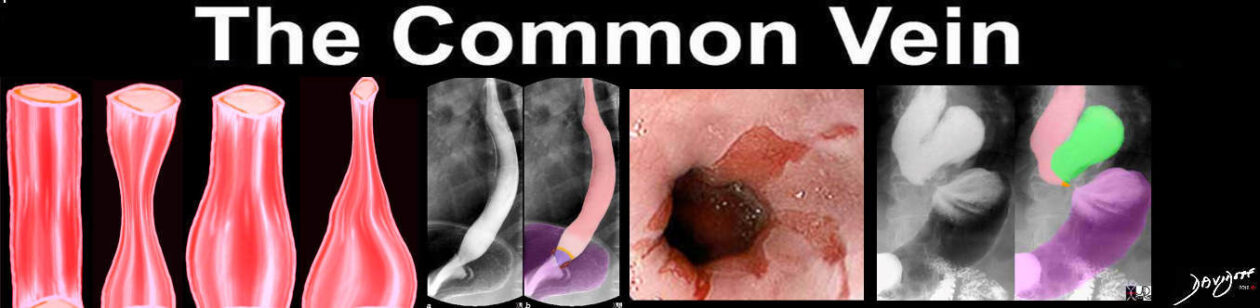
The Esophagus
The Common Vein Copyright 2007
Definition
The esophagus is a tube-like structure that provides a conduit from the mouth and oropharynx to the stomach and it is a key component of the digestive system..
Structurally it is characterized by its muscular and tube like properties. Functionally it acts as a conduit which enables the rapid transport of recently ingested, realatively large food and fluid products from the mouth to the stoamch.. While not essential to life, its inherent properties and function provide a normal mechanism of digestion. Proper coordinated contraction and relaxation , hence propagation, is key to the proper functioning of this very interesting organ.
Many different diseases can affect the esophagus. These range from anatomic alterations within the lumen, to systemic conditions that alter the normal function. These pathologic conditions can cause significant complications ranging from difficulty swallowing to cancer.
Diagnosis of esophageal disease requires careful attention to the deatil of clinical symptoms such as dysphagia, while modalities that include endoscopy, endoscopic ultrasound and barium swallow are essential to diagnosis.
Medical therapy, minimally invasive techniques and surgical options are available depending on the disease process in question.and treatment of the conditions that affect the esophagus.
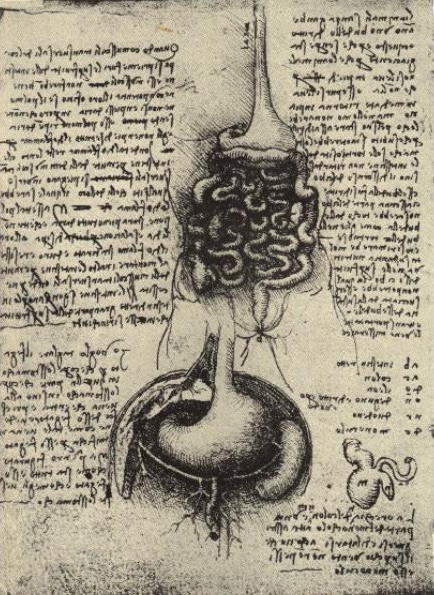 Esophagus and Stomach – da Vinci Esophagus and Stomach – da Vinci |
| 11523 esophagus history da Vinci drawing anatomy historical small bowel Courtesy Leonardo da Vinci |
Principles
If any part of the mucosal, muscular, or neurologic function is not functioning properly, swallowing can be greatly affected. This could lead to dysphagia (problems swallowing), odynophagia (pain with swallowing), or a multitude of processes that will be discussed in this module.
 Esophagus – Air filled Esophagus – Air filled |
| The double contrast barium swallow shows the esophagus as a distended featureless tubular structure with smooth mucosa. This appearance is an artificial appearance of the esophagus since it is as a result of reflux of air from the stomach and inhibition of the primary stripping wave induced by the method of the study.
49741 esophagus normal anatomy double contrast barium swallow upper GI Courtesy Ashley DAvidoff MD |
The unique structural characteristics of the esophagus include its:
- histopathology consisting of squamous mucosa
- striated and smooth musculature
- circular and longitudinal muscles
- ability to change shape to accommodate a swallowed bolus
- lack of a serosal lining (unlike the remainder of the GI system)
The unique functional aspects include its:
- coordinated contraction
- ability to prevent reflux back up into the esophagus
It may be helpful to think of the esophagus as a tube of toothpaste. To expel the toothpaste, one typically squeezes the tube at the end furthest from where the tooth paste is dispensed. If you squeeze from the middle, toothpaste can be expelled in both directions (normally and out the back end). In the esophagus, this could be related to a motility disorder or obstruction from lesions such as rings, webs, or masses.
Again, referring to our toothpaste model, if the tip of the toothpaste dispenser is narrowed from old toothpaste, flow out is reduced. Again, the esophagus can be thought of the same way if there is narrowing at the GE junction expulsion is decreased.
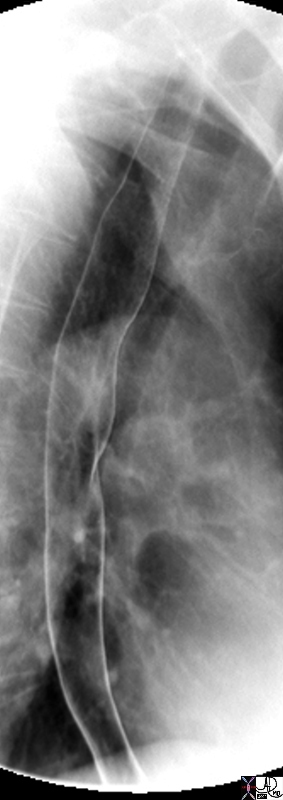
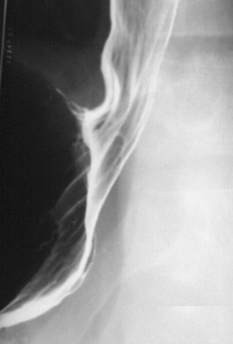
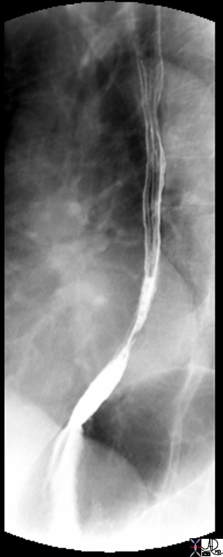 Esophagus – Primary Stripping Wave Esophagus – Primary Stripping Wave |
| This single contrast study shows a relatively decompressed esophagus with the primary stripping wave noted as a constriction in the lower third of the esophagus. The longitudinal folds noted in the upper two thirds of the esophagus are a normal feature of the non distended esophagus.
49743 esophagus normal anatomy double contrast barium swallow upper GI Courtesy Ashley DAvidoff MD |
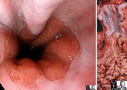
The normal esophagus changes from a whitish hue to a salmon pink at the Z-line. The white color is due to the squamous epithelium of the esophagus and the pink hue is due to the columnar epithelium of the stomach. The image on the left is an endoscopic view and on the right in an anatomical specimen.
Ashley Davidoff, M.D. TheCommonVein.net
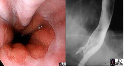
The gastroesophageal junction (also know as the Z line, ZZ line or squamocolumnar junction) is clearly outlined as the squamous epithelium (white) transitions to the gastric epithelium (reddish pink mucosa). It is also exquisitely demonstrated in the double contrast barium swallow as a sharp, contrast and air-filled esophagus and the stomach where no contrast is present.
Courtesy of: Joshua Namias, M.D. and Ashley Davidoff, M.D. TheCommonVein.net
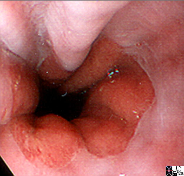
The gastroesophageal junction (aka Z line, ZZ line or squamo-colunar junction) is clearly outlined as the squamous epithelium (white) transitions to the gastric epithelium (reddish pink mucosa)
Ashley Davidoff MD TheCommonVein.net 73474
There are many common disease processes that can be encountered in the esophagus. The most common is gastroesophageal reflux disease (GERD). The reflux of low pH gastric juices thru the normally tight lower esophageal sphincter results in a multitude of complaints. These can range from the classic retrosternal burning to chest pain, water brash, halitosis, to chest pain that simulates angina. Other common conditions encountered include rings/webs, motility disorders, esophagitis, Barrett’s esophagus, and malignancy. These conditions will be discussed in the diseases section below.
Several modalities exist to evaluate a patient’s complaints as they relate to the esophagus. The best study greatly depends on what the diagnostician is looking for. To evaluate the mucosa, examine for luminal lesions, biopsy or treat a ring, an upper endoscopy would be the study of choice.
Radiographic studies are invaluable in the workup of a patient with dysphagia. The barium swallow requires the patient to swallow a combination of barium and effervescent tablets in order to evaluate in real-time, the process of swallowing. This study allows for excellent review of the anatomy and various pathologic processes such as narrowed regions, dilatations, as well as providing an ability to evaluate motility.
CT scan of the chest and abdomen allows for a global view of the esophagus within the mediastinum and chest cavity.
Endoscopic ultrasound (EUS) is an excellent modality that can be used in conjunction with traditional endoscopy to evaluate the structural integrity of the esophagus as well regional lymph nodes. As we discuss specific diseases a more lengthy description of the diagnostic approach will be included.